
 Türkçe

Mole examination is a dermatological evaluation performed to assess the structural features of moles (nevi) on the skin and the possible risk of malignant transformation. During the examination, the physician examines the mole’s color, border, symmetry, and surface features in detail using a dermatoscope device.
Dermatoscopic mole examination plays a critical role especially in the early diagnosis of moles with irregular borders, showing color change, or growing. This method provides high accuracy in the differential diagnosis of melanocytic and non-melanocytic lesions. Early detection is of vital importance in preventing the development of malignant melanoma.
Digital mole follow-up supports the long-term monitoring process of mole examination. Recording moles at regular intervals with computerized imaging systems makes it possible to notice possible changes early. This method increases the effectiveness of routine control in high-risk individuals.
After a mole examination, biopsy or surgical excision may be required if a suspicious lesion is detected. This procedure enables a definitive diagnosis to be made through pathological examination. Regular examinations performed by a specialist dermatologist are recommended in terms of protecting skin health and reducing cancer risk.
| Things You Should Know | Information |
| What is a mole examination? | Mole examination is a dermatological evaluation performed to determine possible risks by assessing the shape, color, size, and border characteristics of moles on the skin. The aim is to distinguish whether the moles are benign or potentially malignant (for example, malignant melanoma). |
| How is the examination performed? | In the physical examination performed by the dermatologist, a special magnification device called a dermoscope is used. With this device, the internal structure of the mole, pigment distribution, vascular structure, and sublayer features are evaluated in detail. |
| When should an examination be performed? | • When a change in color, shape, size, or surface is noticed• When new moles form• In large congenital moles• In moles with irregular borders, multiple colors, or a diameter exceeding 6 mm• In individuals with a family history of skin cancer• Regular examination is recommended for people with fair skin, many moles, or frequent sun exposure. |
| Criteria evaluated | The asymmetry of the mole, border clarity, color distribution, diameter, and changes over time are monitored. In line with these criteria, the risk status of the mole is determined. |
| Path to follow after the examination | Surgical removal and pathological examination may be required for moles with suspicious features. For moles that do not carry risk, control examinations at regular intervals are recommended. |
| Follow-up and precaution | Protection from the sun, regular observation of the skin, and dermatological control at least once a year are important for the early diagnosis of risky moles. Especially in high-risk individuals, follow-up can be performed with digital mole mapping systems. |

Specialist Dr. Seval Karasatı
Dr. Seval Karasatı is a dermatology (skin diseases) specialist in Ankara specializing in skin health, hair and nail diseases, aesthetic dermatology, and laser applications. With her scientifically based approach and aesthetic perspective, she offers personalized treatment plans for each of her patients.
In the field of dermatology, Dr. Seval Karasatı specializes not only in common skin problems such as acne, rosacea, eczema, allergic skin diseases, and hair loss, but also in mole monitoring and the early diagnosis of skin cancers. She provides effective solutions to her patients with modern devices and up-to-date treatment methods.
Serving as a dermatology specialist in Ankara, Dr. Seval Karasatı is also experienced in aesthetic dermatology procedures. She applies holistic treatments aimed at skin renewal and rejuvenation with procedures such as fillers, PRP, mesotherapy, gold needle, and laser treatments.
Dr. Seval Karasatı conducts a comprehensive evaluation for each patient and shapes the treatment process according to personal needs. Her goal is not only to treat diseases but also to help achieve a healthy, vibrant, and aesthetically balanced skin appearance.
Create an Appointment
What Is a Mole (Nevus)?
A mole (nevus) is a generally benign skin formation that may be brown, black, or skin-colored, formed by the gathering of pigment cells called melanocytes in the skin. It may be present at birth or develop over time. Most moles are harmless; however, if there are signs such as changes in shape, color, size, or bleeding, dermatological evaluation is required. Mole follow-up is of great importance in the early diagnosis of skin cancers such as melanoma.
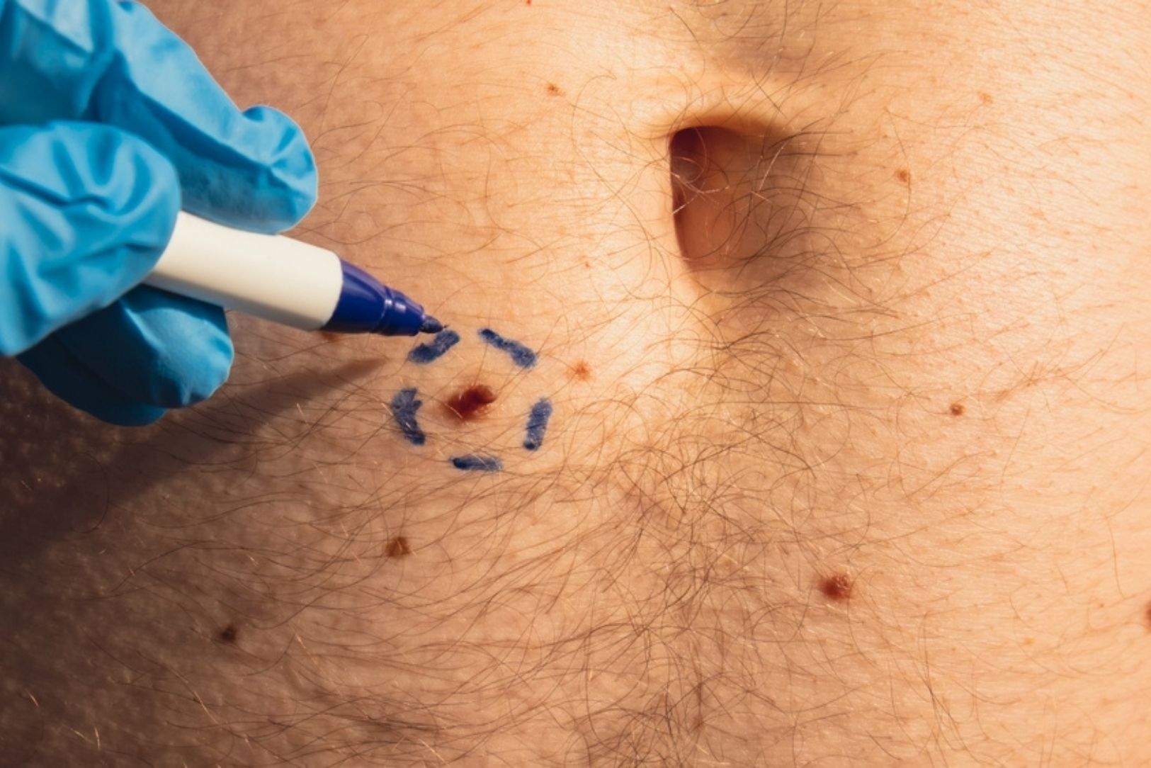
Why is mole examination so important?
The reason this examination is so important can be summarized in one word: Melanoma. The vast majority of moles are completely harmless and remain so for life. However, some moles may have the potential to become cancerous over time, or more importantly, some melanomas may initially look like a normal mole.
If melanoma is noticed while it is still only in the uppermost layer of the skin (at an early stage), it can be completely treated with a simple surgical procedure, and the recovery rate is close to 99%. However, if it is not noticed and progresses to the deeper layers of the skin, it is an extremely aggressive type of cancer with a very high risk of spreading (metastasizing) to other organs of the body (lymph nodes, liver, brain). This is why regular mole examination is the most effective way to catch this dangerous transformation before it becomes a threat.
Who should have regular mole check-ups?
Although everyone should know their skin and consult a doctor in a suspicious situation, some people carry a much higher risk in terms of skin cancer. It is of vital importance for these individuals to be examined regularly by a dermatology specialist, generally once a year, even if they have no complaints.
What are the main risk factors for skin cancer?
When determining a person’s skin cancer risk, we evaluate many factors together. These factors help us determine how often you should come for control and which follow-up methods should be used. The main risk factors are:
- Personal history of skin cancer (melanoma or other types)
- History of melanoma in the family (especially mother, father, sibling)
- A large number of moles on the body (generally more than 50)
- Presence of atypical or unusual (dysplastic) moles
- Fair skin type (skin that burns easily and cannot tan)
- Light-colored eyes (blue, green, hazel)
- Red or blonde hair
- Signs of severe sun damage (excessive freckling, spots)
- Blistering sunburns experienced in childhood or adolescence
- Use of tanning beds (especially at a young age)
- A weakened immune system due to organ transplantation or some diseases
What does having too many moles mean?
People with more than 50 or even more than 100 moles on their body are at a higher risk for melanoma. There are two main reasons for this. The first is that statistically, the more moles you have, the greater the possibility that one of them may become cancerous. The second is a completely logistical problem: among hundreds of moles, noticing one that is newly appearing or changing can be as difficult for both you and your doctor as “finding a tree in the forest.” This condition is called a “high nevus burden,” and advanced technology methods (such as mole mapping) must definitely be used in the follow-up of these patients.
What is an atypical mole (dysplastic nevus)?
Atypical moles are moles that appear “out of the ordinary.” A normal mole is usually small, symmetrical, single-colored, and sharply bordered. Atypical moles are generally:
- Larger (for example: more than 6 mm)
- Irregular and indistinct at the edges
- Asymmetrical (one half does not resemble the other)
They contain more than one color tone (pink, brown, black)
Having one atypical mole does not mean that the mole is “cancer.” However, the presence of such atypical moles on your body is a strong sign showing that your overall risk of developing melanoma is higher than normal. These moles need to be followed especially closely.
How is a head-to-toe skin examination performed?
A head-to-toe skin examination (TBSE) is the systematic examination of the patient’s entire skin surface by a dermatologist. This examination is not just looking at obvious moles. It starts from the scalp and includes every square centimeter of the skin, including the face, behind the ears, neck, trunk, arms, legs, armpits, palms, soles of the feet, between the toes, and even the nail beds. This examination is performed in a well-lit room, usually after you are asked to wear an examination gown. The aim is to create a complete “baseline map” of the current condition of the skin and to compare future changes.
Contact Us for Anything You Wonder About Skin Health!
How should I prepare before coming to the examination?
Some simple preparations are requested from you so that the examination can be as efficient and accurate as possible. These preparations are:
- Coming without makeup (it may hide small lesions on the face)
- Completely removing nail polish (on both fingernails and toenails)
- Leaving the hair open (a bun or ponytail makes examination of the scalp difficult)
Especially having nail polish-free nails is very important, because some extremely dangerous types of melanoma that can develop under the nail bed (acral lentiginous melanoma) can only be noticed when the nails are clean.
How can I monitor my moles myself at home?
The biggest responsibility falls on you between two doctor check-ups. You know your own skin better than anyone else. It is very important that you examine yourself once a month in a well-lit room with the help of a full-length mirror and a hand mirror. Start from your face, carefully examine your scalp (separating it with a comb), behind your ears, the front and back of your trunk (with a hand mirror), your arms, your legs, and especially the soles of your feet and between your toes. You can ask a relative for help for areas you cannot reach (such as your back).
What is the “ABCDE” rule for a suspicious mole?
In your follow-up at home, the “ABCDE” rule will guide you in understanding when you should suspect a mole. It consists of the initials of the five most common features of melanoma. The meanings of these letters are as follows:
- A – Asymmetry (if one half of the mole does not resemble the other half)
- B – Border (border irregularity; if it is indented, protruding, or indistinct)
- C – Color (color diversity; if it is not a single color but contains more than one shade)
- D – Diameter (diameter; generally if it is larger than 6 mm, that is, larger than a pencil eraser)
- E – Evolution / Change (this is the most important rule: the mole growing over time, changing color or shape, becoming raised, itching, or bleeding)
If you notice a mole with one or more of these features, you should consult a dermatologist without delay.
What does the “Ugly Duckling” sign mean?
Although the ABCDE rule is a great guide, sometimes the “Ugly Duckling” sign can be much more life-saving. This concept is based on the following: Most of the moles on a person’s body resemble each other and form a “mole family.” For example, all of a person’s moles may be small, round, and light brown.
The “Ugly Duckling” sign means that the most suspicious mole is the one that does not fit this family, the mole that “does not resemble its siblings.” If all your moles are small and light-colored, but one of them is large, dark-colored, and irregular, that mole is the “ugly duckling” and must definitely be seen by a doctor even if it does not fit the ABCDE rule. This is an important part of an experienced dermatologist’s pattern recognition ability.
What is dermoscopy and why is it used in the examination?
Dermoscopy (or dermatoscopy) is a technology that has revolutionized mole examination. When we look with the naked eye or with a standard magnifier, we cannot see the structures underneath due to the light reflected from the uppermost layer of the skin. By using a special polarized light source and a powerful magnification system (generally 10-40 times), the dermoscope allows us to look “under the hood” of the skin.
Thanks to this device, we can analyze in detail not only the surface of the skin but also the architectural structures in the deeper layers where pigment cells are located, the pigment network, vascular patterns, and color distribution. This is the most important tool that sharpens our “eye” in the early diagnosis of melanoma.
What are the advantages of dermoscopy?
Dermoscopy offers important advantages that greatly increase diagnostic accuracy:
- Reducing unnecessary biopsies (moles that look suspicious but are understood to be benign on dermoscopy)
- Detecting early-stage melanomas (moles that look normal to the naked eye but show suspicious patterns on dermoscopy)
- Diagnosing other skin diseases (hair loss, scabies, fungus)
- Monitoring treatment response (in spot or psoriasis treatments)
- Determining the correct biopsy site (identifying the most dangerous point of a large lesion)
What is digital mole mapping (Total Body Photography)?
This method is a “skin follow-up” system used especially for high-risk patients. Digital mole mapping is the process of taking high-resolution, standardized photographs of the patient’s entire body surface with a special system and uploading them to software. This is, in a way, a “map” of your skin.
This map is generally repeated annually. The aim is to place the two maps on top of each other at the next control and give an objective and definite answer to the questions, “Was this mole here last year?”, “Is there growth in the shape of that mole?”, or “Is this spot completely new?” This is the only reliable way to apply the “E – Evolution” rule, especially in patients with hundreds of moles.
For whom is mole mapping necessary?
This method is not necessary for everyone, but it is of vital importance for the following high-risk patients:
- Those with a large number of moles (generally more than 50-100)
- Those with many atypical (dysplastic) moles
- Those with a personal history of melanoma (for follow-up of a new melanoma or recurrence)
- Those with a strong family history of melanoma (those with genetic predisposition)
What does sequential digital dermoscopy (SDDI) mean?
This is the most advanced method in mole follow-up. This system combines two technologies:
- Total Body Mapping (Macro Image): A general map of your body is created.
- Digital Dermoscopy (Micro Image): Dermoscopic photographs of the moles identified as “suspicious” or “atypical” on the body map are taken one by one and recorded by matching them with their locations on the map.
At your next check-up (for example: 6 months or 1 year later), the dermoscopic images of the same moles are taken again. The software can detect even the slightest structural change that cannot be noticed with the eye by superimposing these two micro images. This is the application of the “Evolution” rule at the microscopic level.
Contact Us for Anything You Wonder About Skin Health!
What do 3D (three-dimensional) mapping systems provide?
While traditional (2D) mapping is based on flat photographs, 3D systems create a three-dimensional “avatar” of your body with special cameras. This provides a great advantage especially in curved areas such as the shoulders, sides of the back, and hips, where 2D photographs may miss or distort areas due to stretching. The 3D model ensures that no point of the skin is overlooked and that the anatomical positions of the moles are recorded much more precisely.
Do these digital follow-up systems prevent unnecessary mole removal?
Absolutely yes. This is one of the greatest benefits of these systems. In the past, the rule of “if in doubt, remove it” was applied for atypical moles in the “gray zone” that looked “a little suspicious” but could not clearly be called “cancer.” This meant a large number of unnecessary surgeries, scars, and anxiety for these patients.
Thanks to digital follow-up systems, instead of removing these “gray zone” moles immediately, we can safely monitor them for 3-6 months. If at the control 6 months later we prove that there has been no change in the digital dermoscopic image of this mole (that is, that it is “stable”), we obtain very strong evidence that the mole is benign. This transforms the rule of ‘if in doubt, remove it’ into the rule of ‘if you are not sure, follow it,’ protecting the patient from unnecessary surgery.
How is it decided that a mole is suspicious?
The decision to take a biopsy (sample) from a mole is a combination of all these steps. While making the decision, the patient’s history (is the mole changing?), clinical examination (ABCDE/Ugly Duckling), and dermoscopic examination (suspicion of cancer in the internal structure) are evaluated together. If a lesion raises serious suspicion in any of these filters, a tissue sample must be taken for a definitive diagnosis.
How is a biopsy (sample) taken from a suspicious mole?
When suspicion of melanoma is high, the “gold standard” biopsy method is “excisional biopsy.” This means the entire mole is surgically removed under local anesthesia, together with a small rim of healthy skin tissue of 1-3 mm around it, including all layers of the skin, and sent to pathology. This is a minor surgical procedure requiring stitches.
Why is it so important to remove the entire mole?
In the pathology laboratory, when a diagnosis of melanoma is made, the most important thing the pathologist must examine is the “Breslow Depth.” This is a millimetric measurement showing how deeply this cancer has gone from the uppermost layer of the skin. The patient’s entire future treatment plan and prognosis (the course of the disease) are determined according to this depth.
If only a small piece is taken from a suspicious mole by “shaving” or “punch,” there is a risk of missing the deepest part of the lesion. According to the piece in hand, the pathologist may say “0.5 mm deep,” but in the part that was not taken, the lesion may perhaps have reached a depth of 2 mm. This can be a very serious mistake that will misdirect the patient’s treatment (incomplete staging). Therefore, in order to preserve diagnostic integrity and make the most accurate staging, it is preferred to remove the entire suspicious lesion in one piece.
What happens if the biopsy result comes back as “melanoma”?
Although this is a frightening moment, it is the first step of treatment when detected early. If the result comes back as melanoma, the first thing to do is to look at the Breslow depth in the pathology report. Depending on this depth, a second surgical procedure (wide excision) is planned in accordance with international guidelines. In this procedure, depending on the depth of the cancer, the area around the first biopsy site is removed again with a wider safety margin. If necessary, an additional procedure (sentinel lymph node biopsy) may be performed to understand whether the cancer has spread to the lymph nodes. The basis and primary cure of melanoma treatment is surgery.
In summary, what should be considered in mole examination and follow-up?
The follow-up of your moles is a joint effort between you (self-examination at home) and your dermatologist (professional examination). Knowing your risk profile (fair skin, many moles, family history) and acting accordingly is the first step. Regularly checking your own skin according to the ABCDE rule and the Ugly Duckling sign is your earliest warning system.
In professional examination, dermoscopy strengthens our “eye,” allowing us to see details that the naked eye cannot see. If you are in a high-risk group, digital mole mapping and sequential dermoscopy protect you from both unnecessary surgeries and catch a real danger at the earliest possible moment by monitoring changes objectively.
Remember, melanoma is a type of cancer that can be treated at a rate close to 100%; as long as it is diagnosed early. Mole examination is the key to this early diagnosis.
Frequently Asked Questions
Why is mole examination performed and in which situations is it necessary?
Mole examination is performed to evaluate whether moles on the skin carry a risk of becoming cancerous. Dermatological evaluation is essential especially for moles that grow rapidly, change shape and color, itch, or bleed.
What is the use of dermatoscopy in mole examination?
Dermatoscopy allows the microscopic structure of the mole to be examined by magnifying it. Thanks to this method, the structure, color distribution, and border features of the mole are evaluated, and the possibility of malignant melanoma is detected at an early stage.
How often should a mole examination be performed?
People with fair skin, many moles, or a family history of skin cancer should have a mole examination at least once a year. In high-risk individuals, this period can be shortened to six months.
What should be done when a change is noticed in moles?
If darkening in the color of a mole, asymmetry, border irregularity, or sudden growth is noticed, a dermatologist should be consulted without delay. Early diagnosis significantly increases the success rate in skin cancer treatment.
Should mole examination also be performed in children?
Yes, congenital or later-developing moles in children should also be monitored. Especially moles in areas prone to trauma should be checked regularly, and dermatoscopic evaluation should be performed if necessary.
Why is it important to perform a mole examination before mole removal?
Examination before mole removal is necessary for the procedure to be performed safely. While moles that carry a cancer risk need to be removed surgically, harmless ones can be removed with laser or radiofrequency.
What happens if a suspicious lesion is detected as a result of a mole examination?
If a suspicious lesion is detected, the dermatologist usually recommends a biopsy. If cancerous transformation is detected in the cells as a result of the biopsy, the disease can be brought under control early by planning appropriate surgical treatment.
What should be considered before a mole examination?
Before the examination, the skin should be clean, and makeup or colored cream should not be used. If there is sunburn, the appointment may be postponed because color changes in the skin may make evaluation difficult.
Why is a dermatological examination essential before mole removal with laser?
Laser treatment can only be safely applied to harmless moles. Therefore, performing a dermatological examination beforehand to determine the type and risk status of the mole prevents possible complications.
How does mole examination play a role in the early diagnosis of skin cancer?
Mole examination enables skin cancer to be noticed at the earliest stage. Thanks to dermatoscopic analysis, malignant changes can be detected before they show symptoms, and treatment is started early.
How Long Does a Mole Examination Take?
A mole examination usually takes between 10 and 30 minutes. The dermatologist examines the moles in detail with a special device called a dermatoscope. If there are many moles on the body, the screening time may be longer. If a suspicious lesion is detected, an additional procedure may be planned for biopsy. Although the duration of routine check-ups is short, it is important to allow time for a detailed evaluation.
Which Department or Doctor Should Be Consulted for a Mole Examination?
For a mole (nevus) examination, the dermatology department should be consulted. Dermatologists examine moles in detail with methods such as dermatoscopy and can provide referral for biopsy or surgical removal when necessary.
How is a mole examination performed?
Mole examination is performed by a dermatology specialist with the naked eye and with a magnifying device called a dermatoscope. The color, shape, borders, and symmetry of the moles are evaluated, and suspicious lesions are detected at an early stage.
Why is mole screening performed?
Mole screening is performed to detect the risk of skin cancer and especially melanoma at an early stage. Regular screenings enable dangerous changes in moles to be noticed and treated in a timely manner.
How is self mole examination performed?
Self mole examination is performed once a month by checking the entire body with the help of a mirror. If growth, color change, irregular border, or bleeding is noticed in moles, a dermatology specialist should be consulted.