
 Türkçe

Mole removal is the procedure of safely removing pigmented or raised lesions on the skin using medical methods. The procedure can be performed using laser, surgical excision, or electrocautery methods. Which technique will be chosen is determined according to the structure of the mole, its size, and the skin type.
Laser mole removal is especially preferred for small, superficial moles on the face and neck area. Laser beams target the pigment cells and vaporize the mole. Rapid healing is achieved after the procedure, and the risk of scarring is quite low.
Surgical mole removal is applied for deeply located moles or moles suspected of cancer. In this procedure, performed under local anesthesia, the mole is completely removed and stitches are placed if necessary. The removed tissue is sent for pathological examination and a definitive diagnosis is established.
In the period after mole removal, it is important to keep the area clean and protect it from direct sunlight. The scab should not be picked, and the healing creams recommended by the doctor should be used regularly. In procedures performed with the correct technique, both the aesthetic appearance and skin health are preserved.
| Things You Should Know | Information |
| What is a mole (nevus)? | They are lesions that form as a result of the accumulation of pigment cells called melanocytes in the skin, usually brown or black in color, with a flat or raised appearance. |
| When is mole removal necessary? | If the mole changes in shape, size, or color; if its borders become irregular; if itching, bleeding, or crusting develops; or if it causes aesthetic or functional discomfort, it may need to be removed. |
| How is mole removal performed? | It is performed under local anesthesia by removing the mole together with some normal tissue around it using a scalpel. The most commonly used method is “elliptical excision.” |
| Alternative methods | For some benign and superficial moles, laser, cryotherapy (freezing), electrocautery (burning), or shave excision methods may be preferred. |
| Post-procedure care | The treated area should be kept clean, dressings should be applied as recommended, and trauma should not be applied to the area. Sun exposure should be avoided, and if there are stitches, they should be removed within the period specified by the physician. |
| Recovery process | Although it varies depending on skin type, the size of the mole, and the width of the removed area, healing is generally completed within a few weeks. A stitch mark may form. |
| Risks and things to consider | Situations such as scarring, infection, bleeding, recurrence of the mole, and failure to completely remove deeply located cells may occur. Suspicious moles must definitely be sent for pathological examination. |

Specialist Dr. Seval Karasatı
Dr. Seval Karasatı is a dermatology (skin diseases) specialist in Ankara specializing in skin health, hair and nail diseases, aesthetic dermatology, and laser applications. With her scientifically based approach and aesthetic perspective, she offers personalized treatment plans for each of her patients.
In the field of dermatology, Dr. Seval Karasatı specializes not only in common skin problems such as acne, rosacea, eczema, allergic skin diseases, and hair loss, but also in mole monitoring and the early diagnosis of skin cancers. She provides effective solutions to her patients with modern devices and up-to-date treatment methods.
Serving as a dermatology specialist in Ankara, Dr. Seval Karasatı is also experienced in aesthetic dermatology procedures. She applies holistic treatments aimed at skin renewal and rejuvenation with procedures such as fillers, PRP, mesotherapy, gold needle, and laser treatments.
Dr. Seval Karasatı conducts a comprehensive evaluation for each patient and shapes the treatment process according to personal needs. Her goal is not only to treat diseases but also to help achieve a healthy, vibrant, and aesthetically balanced skin appearance.
Create an Appointment
Why is the decision made to remove a mole?
There are basically two main reasons for removing moles: health and aesthetics. The most mandatory reason in terms of health is the suspicion that a mole may turn into a “malignant” structure, in other words, the risk of cancer. If suspicious features are seen in the dermoscopic examination (a special illuminated magnifier) performed by your dermatologist, that mole must be removed for a definitive diagnosis.
The other reason is entirely cosmetic or functional. A person may be disturbed by the appearance of the mole. Or the mole may be located in an area where clothing or jewelry is constantly worn, rubbed, bleeds, or becomes irritated. In such cases, even if there is no suspicion of cancer, removing the mole may be preferred to improve quality of life.
Is every mole removed with the same method?
No, the structure and depth of every mole are different, so the method to be applied also changes. The classification of the mole determines how deep the intervention needs to go.
More superficial, brown spot-like (junctional) moles can sometimes be treated with laser systems (Q-switched or normal mode pigment lasers).
Deeper, raised, skin tag-like (dermal or compound) moles generally require methods that go deeper.
For these deep moles, either full-thickness surgical removal (excision) or ablative lasers (Carbon Dioxide or Erbium) that resurface the skin may be used. If surgical excision is chosen, reaching the correct depth so that no part of the mole remains behind is a critical step that depends on the experience of the physician performing the procedure.
How can it be understood whether a suspicious mole is dangerous?
The definite and only way to determine whether a mole is dangerous is to remove that mole and examine it in the pathology laboratory. The method of taking a biopsy from a mole suspected of melanoma (the most dangerous type of skin cancer) is a vital decision that directly affects the patient’s entire treatment process and the course of the disease (prognosis).
If melanoma is suspected, the gold standard method is excisional biopsy. This means removing the entire suspicious mole together with some healthy tissue around it, including all layers of the skin (epidermis, dermis, and part of the fatty tissue beneath it). This method provides the pathologist with the most accurate and complete information for diagnosis.
Why is only a small part of a suspicious mole not taken?
Because this may lead to an “incomplete” or “incorrect” diagnosis. Sometimes only a part of the mole is taken with a “punch” biopsy or a superficial “shave” biopsy. However, this incomplete sampling carries serious risks.
The most important thing the pathologist needs to know in order to determine the stage of the cancer is the “Breslow depth,” that is, the thickness of the tumor. If only a partial sample is taken, this thickness may be measured as less than it actually is. For example, if the thickest part of the melanoma is 2 mm but the sample taken shows only a 0.5 mm section, the treatment plan will be completely wrong.
Studies have shown that in 18% of patients diagnosed with a partial biopsy, treatment plans had to be changed after the entire mole was removed. In contrast, this rate is only 2% in patients whose entire mole was removed from the beginning (excisional biopsy). Therefore, if there is any suspicion, removing the entire mole is the safest way to prevent delay and failure in treatment.
What does an “atypical” (Dysplastic) mole mean and is another surgery required?
“Atypical” or “dysplastic” moles are moles that are not cancerous but appear somewhat more irregular than normal moles. If this result appears in your pathology report, it does not mean that you have cancer.
The management of these moles depends on the details in the pathology report:
If the mole is “mildly” or “moderately” atypical and the surgical margins are “clear” (that is, the entire mole has been removed), usually no additional procedure is required.
If the surgical margins are “positive” (that is, atypical cells remain at the cut edge), the situation changes. In mildly atypical moles, if there is no visible residue (pigment), follow-up alone may be sufficient. In moderately atypical moles, options such as re-excision of the remaining tissue or very close follow-up are evaluated. This decision is made entirely according to the clinical condition.
What preparations are made before the mole removal procedure?
Pre-procedure preparation is important both for the success of the procedure and for your safety. During this process, the following are done:
- Photography: Before the procedure, photographs of the mole are definitely taken from more than one angle to record its current condition. This is both a legal requirement and a reference point for future follow-ups.
- Consent Form: Your doctor explains the procedure to be performed, its alternatives, its risks (such as infection, bleeding, scarring, wound opening), and what may happen if the procedure is not performed. You are asked to sign an informed consent form stating that you understand this information.
- Medical History Inquiry: It is questioned whether you have any condition that especially affects blood clotting or impairs wound healing (for example diabetes, smoking).
Is it necessary to stop using medication before the procedure?
Yes, this is very important. Some medications and supplements that increase the risk of bleeding need to be discontinued a certain period before the procedure (usually 3-7 days). The following are things you should definitely consult your doctor about:
- Aspirin
- Other blood-thinning medications (Coumadin, Plavix, Xarelto, etc.)
- Some painkillers (NSAID group such as Ibuprofen, Naproxen)
- Vitamin E
- Fish oil (Omega-3)
- Ginkgo biloba
- Herbal supplements such as garlic tablets
In addition, it is strongly recommended to stop smoking for a period before the procedure, as it seriously impairs wound healing.
Is pain felt during the mole removal procedure?
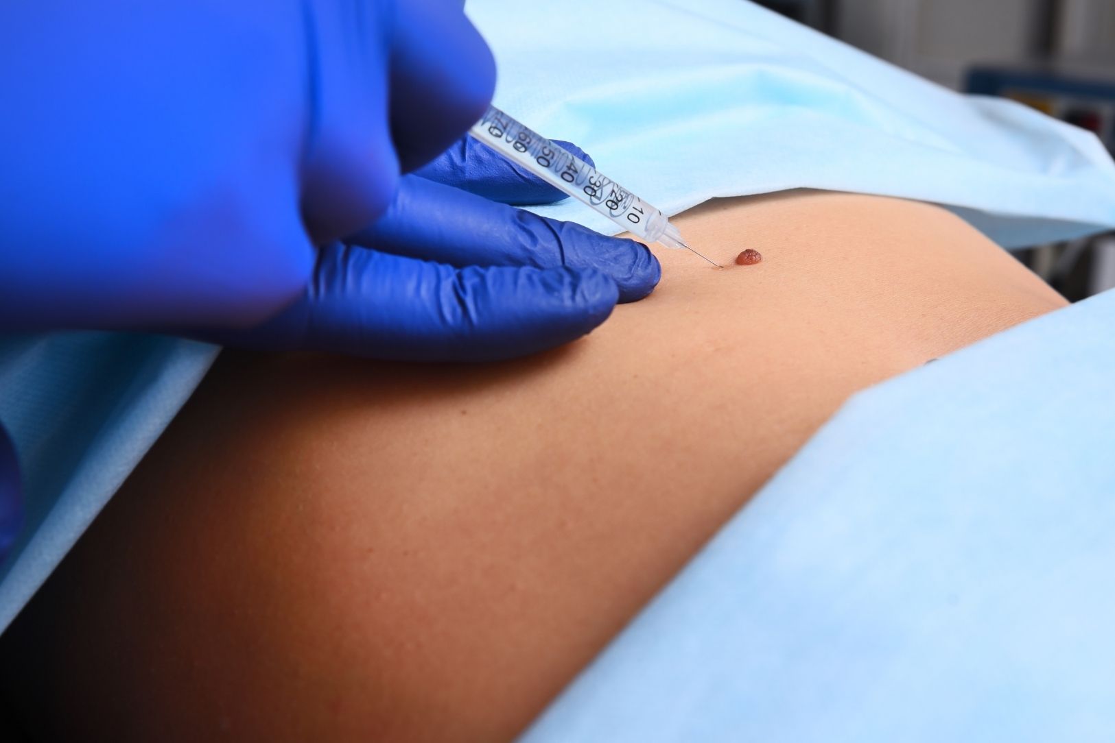
No. The entire procedure is performed under local anesthesia. This only means numbing the area where the mole is located.
The procedure begins with a small injection into the area. Local anesthetic medications containing “epinephrine” are generally used. Epinephrine constricts the vessels in the area, greatly reducing bleeding and allowing the numbness to last longer. After the injection, 3-5 minutes are waited for the drug to take full effect and constrict the vessels. This waiting period is critically important for making the procedure more comfortable and bloodless. When the procedure begins, you do not feel any pain or ache, only a sensation of touch may be present.
How is surgical mole removal performed?
The most commonly used method and the one accepted as the gold standard is “elliptical excision,” that is, removal in a diamond-slice shape.
The purpose of this is to prevent bulges called “dog ears” from forming at the ends while bringing the wound edges together after the mole is removed. The direction of this incision is also very important. The incision is planned parallel to the natural tension lines of the skin (RSTL or Kraissl lines). You can think of them like the expression lines on your face or the lines that form when you bend your elbow. If the incision is made parallel to these lines, there is minimal tension while the wound heals and the remaining scar becomes much less noticeable.
In this technique, the standard geometry is for the incision length to be 3 times its width (3:1 ratio). This ratio ensures the most aesthetic closure of the wound.
Contact Us for Anything You Wonder About Skin Health!
What does it mean to leave a “safety margin”?
Removing some healthy tissue around the mole together with the mole itself is called a “safety margin” or “surgical margin.”
If the removed mole is benign, this margin is enough to allow tension-free closure of the wound (for example 1-2 mm).
However, if a suspicious mole or a diagnosed melanoma is being removed, these margins are determined by international guidelines and are kept much wider. The aim is also to clear “subclinical” cancer extensions that are not visible to the naked eye but may be present under the microscope. These margins vary according to the thickness of the mole measured in pathology (Breslow depth).
- Melanoma in situ (earliest stage)
- Melanoma with a thickness less than 1 mm
- Melanoma with a thickness between 1-4 mm
- Melanoma with a thickness greater than 4 mm
The millimetric or centimetric safety margins that must be left are different for each.
Are there stitch-free methods for benign moles?
Yes, if it has been established with certainty through clinical and dermoscopic examination that a mole is benign, priority may be given to the aesthetic result. In such cases, methods other than traditional stitched surgery may be preferred.
- Shave Excision: It is especially used in areas such as the face where there is a risk of raised scarring or in superficial, raised moles. The mole is cut horizontally at skin level. Since the lower layer of the skin (dermis) is preserved, stitches are generally not required and the risk of scarring is lower.
- Punch Excision (Removal with a Punch): It is especially ideal for small, dome-shaped moles on the face. Instead of a normal diamond-slice incision, the mole is removed with a round “punch” appropriate to the size of the mole. Since the removed tissue is very small, it is usually closed with a single stitch or sometimes left to heal without stitches. Since less healthy tissue is removed, the scar is very small.
- Laser Mole Treatment: As mentioned above, lasers targeting pigment or resurfacing the skin are also an option for suitable superficial moles.
How is bleeding stopped during the procedure?
Keeping bleeding under control during the procedure (hemostasis) is very important both for the surgeon to work comfortably and for post-procedure wound healing. The epinephrine given by injection already greatly reduces bleeding from the beginning by constricting the vessels.
Despite this, if small bleedings occur, different methods are used:
- Direct Pressure: Applying pressure to the area with a swab is usually sufficient.
- Electrosurgery (Cautery): This is the most commonly used method. Bleeding is stopped instantly by touching small vessel points with a fine tip.
- Ligation with Sutures: Rarely, if a larger vessel bleeds, this vessel can be tied with a surgical thread.
- Chemical Agents: In superficial oozing, special solutions that stop bleeding (such as aluminum chloride) can be applied.
How are stitches placed to achieve the best scar?
The secret of successful wound closure is managing the tension on the wound edges. The more tension there is on the wound, the more it scars.
To prevent this, a layered suturing technique is used. This does not simply mean stitching the wound only from the surface.
- Deep Sutures: First, absorbable sutures are placed in the lower layers of the skin (dermis and subcutaneous tissue). The purpose of these sutures is to carry the main load of the wound and reduce the surface tension to zero. These sutures are not visible from the outside and do not need to be removed.
- Surface Sutures: After the deep sutures have taken the tension, the uppermost layer of the skin (epidermis) is closed edge to edge with very fine threads. The purpose of these sutures is only alignment.
These surface sutures are removed between 4 and 14 days depending on the area. For example, while 4-6 days are sufficient on the face, in more tensioned and slower-healing areas such as the back or leg, the sutures may need to remain for 14 days.
Why is the removed mole sent to pathology?
No matter how “benign” it appears to the eye, every surgically removed mole must definitely be sent to the pathology laboratory. This is a medical necessity. Sometimes atypical cells at the microscopic level or an early-stage melanoma may be hidden under a completely harmless-looking mole.
The pathology report does not only tell us whether the mole is benign or malignant. At the same time, it confirms whether the surgical margins are clear (whether the mole has been completely removed) and, if it is malignant, its thickness (Breslow). The next step of the treatment is planned according to this report.
How is the removed tissue preserved so that the pathology result is accurate?
The accuracy of the pathology report depends not only on the pathologist but also on the period from the moment the mole is removed until it enters the laboratory. The tissue begins to deteriorate the moment it is separated from blood circulation.
To stop this deterioration, the tissue must be placed in a preservative liquid called a “fixative” (usually formalin). There are two critical rules in this process:
Warm Ischemia Time: The time between removal of the mole and placement in formalin should ideally be less than 20 minutes. As the time lengthens, proteins in the tissue break down and this disrupts the special staining (immunostaining) tests that the pathologist will perform.
Thickness Limit: Formalin penetrates tissue very slowly (approximately 1 mm per hour). If the removed piece is too thick (for example thicker than 2 mm), it takes hours for the liquid to reach the center of the tissue. During this time, while the outside of the tissue is being fixed (preserved), the inside deteriorates. Therefore, thick pieces must be sliced by the surgeon before being sent to pathology for the reliability of the result.
What are the risks of the mole removal procedure?
Mole removal is generally a very safe and low-risk procedure. However, as with every surgical intervention, there are some possible complications:
- Wound infection
- Bleeding or hematoma (accumulation of blood under the tissue)
- Suture reaction (allergy or inflammation against the stitches)
- Delayed wound healing
- Wound opening (stitch separation)
- Hypertrophic scar (raised scar)
- Keloid (abnormal growth of the scar)
- Recurrence in the mole (if it cannot be completely removed)
What factors increase the risk of wound opening (stitch separation)?
Wound opening (surgical wound dehiscence) is one of the most important complications that impair healing. Some conditions increase this risk:
- Advanced age (80 years and older)
- The wound being in very mobile or tense areas such as the lower parts of the legs
- Bleeding or infection developing after the procedure
- Smoking (because it impairs blood flow to the tissue)
It should especially not be forgotten that post-procedure bleeding is a strong risk factor for wound opening. Therefore, if bleeding occurs after the procedure, it must definitely be managed as the doctor instructed (by pressing continuously with a clean cloth for 20 minutes).
Contact Us for Anything You Wonder About Skin Health!
How should wound care be done at home after the procedure?
Good wound care prevents infection and provides a better cosmetic result. The basic rules are as follows:
- For the first 24-48 hours, the wound is generally kept dry and covered.
- After this period, the wound can be gently washed 1-2 times a day with clean water and a mild soap as recommended by your doctor.
- The most important rule: The wound must be kept moist. Scabbing slows healing.
- After washing, the wound should be gently dried and a thin layer of antibiotic ointment or (more commonly) petroleum jelly should be applied on it.
- It should be covered with a non-stick dressing.
Until the wound heals, solutions such as alcohol, tincture of iodine, or oxygenated water (hydrogen peroxide) should DEFINITELY not be used. These products damage the new cells that provide healing and delay the process.
Until the stitches are removed, strenuous and stretching movements (such as heavy sports) that strain the area should be avoided.
What can be done to minimize the stitch mark?
A scar is part of the body’s natural healing process and cannot be completely eliminated, but its appearance can be improved. Scars are initially red and raised, then fade and soften over the months.
What should be done to optimize the scar:
- Protection from the Sun: This is the most critical rule. Even after the stitches are removed, the scar should be protected from the sun for at least 6 months to 1 year. Scars exposed to UV light during the healing period develop discoloration (pigmentation) and leave a permanent brown stain. High-factor sunscreen creams or dressings should be used.
- Moisturization: Keeping the area continuously moist (with petroleum jelly or special scar creams) regulates collagen production.
- Silicone Sheets/Gels: Silicone-based products recommended by your doctor are very effective, especially in preventing the formation of raised scars (hypertrophic scars).
- Massage: Gentle massages to the area after the wound closes can soften the tissue.
- Not Scratching: The wound may itch while healing. Scratching the area or picking the scabs permanently worsens the scar.
Is follow-up necessary after a mole is removed?
Yes. Even if the pathology result is benign, especially people with many moles or a family history of skin cancer should continue annual dermatologist follow-up. The fact that one mole has been removed does not mean that a new suspicious mole will not appear elsewhere. Early diagnosis is life-saving in skin cancers.
Frequently Asked Questions
In which cases is mole removal (excision) preferred?
Mole removal (excision) is preferred when the mole carries a risk of cancer, changes shape, or becomes irritated. It is also applied for aesthetic reasons or for removing moles that are constantly exposed to friction.
Does mole removal by the excision method leave a permanent scar?
After mole removal by excision, a small surgical scar may remain. However, when stitches are placed with aesthetic techniques and wound care is done correctly, the scars lose their prominence over time and adapt to the skin tone.
How long does the recovery process take after mole removal?
Healing after excision is generally completed within 7–14 days. However, full skin renewal may take several weeks. During this process, it is important to keep the area clean, protect it from the sun, and follow the doctor’s recommendations.
Is stitch removal necessary after mole removal excision?
Yes, in the classical excision procedure, non-dissolving stitches are generally used and they are removed after 7-14 days. However, in some cases, dissolving stitches are preferred, in which case stitch removal is not necessary.
Why is mole removal excision important in terms of cancer risk?
Excision allows the removed mole to be sent for pathological examination. In this way, it is determined whether the mole is benign or a precancerous lesion. Early diagnosis plays a critical role in reducing the risk of skin cancer.
Can the mole removal procedure be performed with local anesthesia?
Yes, the excision procedure is generally performed under local anesthesia. In this way, the patient does not feel pain throughout the procedure, the intervention is completed comfortably, and the patient can return to daily life on the same day.
Is there a risk of the mole appearing again after mole removal?
When the root of the mole is completely removed with full-thickness excision, the risk of recurrence is very low. However, if cell residue remains in superficially removed moles, regrowth may be seen, in which case reevaluation is necessary.
Can mole removal excision be performed on the face area?
Yes, moles in the face area can also be removed by excision. In this case, aesthetic suturing techniques are used and the incisions are made parallel to the skin lines in order to keep scars to a minimum.
How should wound care be done after mole removal?
After the procedure, the area should be kept clean and the cream or ointment recommended by the doctor should be used regularly. Not picking the wound during the scabbing period and protecting it from the sun accelerate healing and reduce the risk of scarring.
When is the pathology result available after mole removal excision?
The pathological examination result of the removed mole is generally available within 5–7 days. This report provides definitive information about the structure of the mole and guides additional treatment planning when necessary.