
 Türkçe

Dermatological examination is a medical evaluation process that includes the detailed examination of the skin, hair, nails, and mucosal surfaces. This examination is of great importance for the early diagnosis, classification, and creation of an appropriate treatment plan for skin diseases. Visual observation, palpation, and dermatoscopic examination are the basic steps.
A comprehensive dermatological evaluation begins with taking the patient’s history in detail. The onset time, duration, and triggering factors of complaints such as itching, rash, spots, or masses are questioned. In addition, medications used, allergy history, and systemic diseases are also taken into consideration in the diagnostic process.
During the physical examination stage, the morphology, distribution, and color of lesions are evaluated. If necessary, the dermatologist benefits from additional diagnostic methods such as dermatoscopy or biopsy. In this way, a distinction can be made between benign and malignant skin lesions, and diagnostic accuracy is increased.
The findings obtained after dermatological examination make it possible to create a personalized treatment plan. When deemed necessary, the diagnosis is supported by laboratory tests, allergy evaluations, or imaging methods. Regular dermatological check-ups are of critical importance for the protection of skin health and early diagnosis.
| Things You Should Know | Information |
| What is a dermatological examination? | Dermatological examination is the process of observing and evaluating the skin, hair, nails, and mucosa by a dermatologist. The aim is the diagnosis of existing complaints, the early detection of skin diseases, and the determination of an appropriate treatment plan. |
| How does the examination process begin? | Before the examination, the patient’s detailed medical history is taken. Past skin diseases, the onset time and course of the complaints, products used, allergy history, and whether there is a family history of skin disease are questioned. |
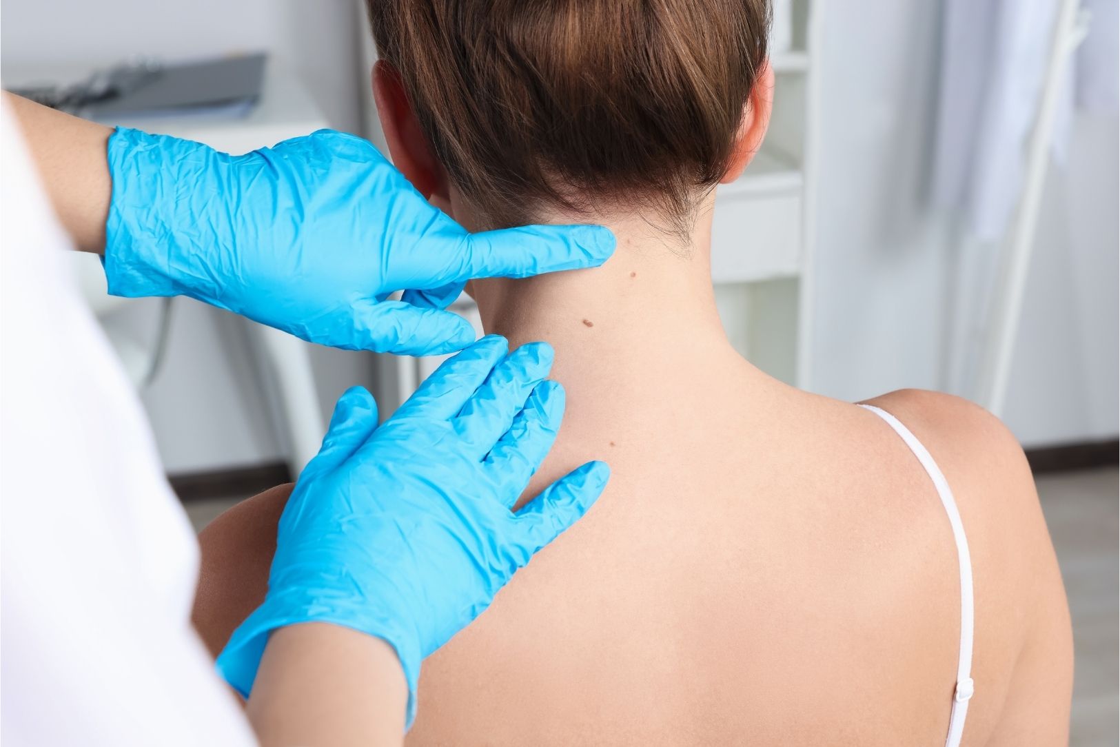
| How is the physical examination performed? | The dermatologist systematically examines the patient’s skin. The face, scalp, trunk, extremities, nails, and, if necessary, mucosa are evaluated visually and, if necessary, by palpation. |
| Auxiliary diagnostic tools | When deemed necessary, moles or lesions are examined in more detail with a magnifying device called a dermatoscope. In addition, in some cases, biopsy, blood tests, fungal tests, allergy tests, or imaging methods may be required. |
| Purpose and importance | Early diagnosis of many diseases, including skin cancer, can be achieved. Chronic skin diseases are monitored, and aesthetic or functional skin problems are evaluated. Personalized care and treatment recommendations are created. |
| When should a dermatologist be consulted? | If there are findings such as color change, spots, moles, itching, redness, wounds, rash, hair loss, or nail changes on the skin; if growth, color change, or border irregularity is noticed in moles; in chronic skin problems or for annual check-up purposes, a dermatologist should be consulted. |

Specialist Dr. Seval Karasatı
Dr. Seval Karasatı is a dermatology (skin diseases) specialist in Ankara specializing in skin health, hair and nail diseases, aesthetic dermatology, and laser applications. With her scientifically based approach and aesthetic perspective, she offers personalized treatment plans for each of her patients.
In the field of dermatology, Dr. Seval Karasatı specializes not only in common skin problems such as acne, rosacea, eczema, allergic skin diseases, and hair loss, but also in mole monitoring and the early diagnosis of skin cancers. She provides effective solutions to her patients with modern devices and up-to-date treatment methods.
Serving as a dermatology specialist in Ankara, Dr. Seval Karasatı is also experienced in aesthetic dermatology procedures. She applies holistic treatments aimed at skin renewal and rejuvenation with procedures such as fillers, PRP, mesotherapy, gold needle, and laser treatments.
Dr. Seval Karasatı conducts a comprehensive evaluation for each patient and shapes the treatment process according to personal needs. Her goal is not only to treat diseases but also to help achieve a healthy, vibrant, and aesthetically balanced skin appearance.
Create an Appointment
Why Does a Dermatological Examination Begin with a Detailed Conversation?
The foundation of the dermatological examination is the detailed medical history taken from the patient, called ‘anamnesis.’ This is similar to putting together the pieces of a puzzle. The diagnostic process begins with understanding the real motivation behind the patient’s visit. It is necessary to meticulously document what the main complaint is, such as itching, a specific lesion, hair loss, or nail change, and in what order the symptoms appeared.
Why Are the Timing and Sequence of Complaints So Important?
The relationship between itching and visible skin lesions is a critical filter that guides diagnosis. The clinician needs to clarify which symptom appeared first: Did the itching start before the lesions, or did the lesions and itching appear at the same time? The answer to this simple question helps narrow down the possible causes.
- Itching First: If itching begins when there are no visible skin lesions and the lesions develop later due to scratching, this generally raises suspicion of an allergic basis (for example atopic dermatitis) or an internal cause (a systemic disease).
- Lesion First: Conversely, if the lesions appear first and itching starts with them or immediately afterward, parasitic (for example scabies) or infectious (for example fungal) causes are considered first.
What Information About the Onset and Progression of the Disease Is Valuable?
When, where on the body, and how the disease began, its duration, and its subsequent evolution (healing, worsening, spreading) should be comprehensively documented. The patient’s age at onset is also of great importance: while symptoms appearing in a young patient may indicate a more allergic basis, similar complaints beginning in an older adult may suggest an underlying systemic, metabolic, or autoimmune disorder, or even a skin neoplasm (cancer).
In addition, knowing whether disease activity is related to seasonality (for example increasing during pollen season) may bring environmental allergies to mind. The patient’s response (or lack of response) to previously tried treatments provides both diagnostic clues and critical information for the future treatment plan.
How Do the Medications You Use and Your Allergies Affect the Diagnosis?
A detailed pharmacological (medication) history inquiry is mandatory. This includes not only current and past prescription medications but also the following.
- Over-the-counter medications
- Vitamins and minerals
- Herbal supplements
Topical (applied) treatments (Especially the strength and amount of use of steroid creams)
The allergy history should include environmental sensitivities as well as known drug reactions.
- Nickel or other metals
- Perfumes and fragrances
- Latex
- Insects
- Pets
- Plants
To understand the patient’s atopic background (allergic predisposition), past allergic disorders such as childhood eczema, asthma, and hay fever must definitely be documented. Lifestyle factors such as home conditions (dampness), social habits (smoking), and occupational exposure further clarify the clinical picture.
Why Are Other Parts of Your Body Questioned Besides Skin Complaints?
Findings on the skin can often be the outward manifestation, that is, an “indicator,” of an internal or systemic disease. For this reason, a “Review of Systems” (ROS) covering all body systems is a fundamental part of the dermatological examination. While this inquiry includes other symptoms related to the skin (changes in moles, rashes, hives), its main purpose is to see the broader underlying picture.
What Does the Review of Systems (ROS) Inquiry Cover?
This inquiry covers the whole body, starting from general symptoms. The main systems and symptoms questioned are:
- General health (Fever, fatigue, weight loss)
- Heart and lung complaints
- Gastrointestinal problems
- Neurological symptoms
- Musculoskeletal pain
- Hematological (Easy bruising, bleeding)
- Immunological (Food or seasonal allergies)
Especially symptoms such as easy bleeding, lymphedema (swollen lymph nodes), or allergies may be clues to systemic diseases presenting with skin symptoms.
What Is a Total Body Skin Examination (TBSE) and Why Is It Necessary?
A Total Body Skin Examination (TBSE) is the cornerstone of dermatological preventive care and requires meticulous, systematic evaluation of the entire skin system from head to toe. The primary purpose of this examination is to ensure the early diagnosis of skin cancers, especially melanoma, which is considered the deadliest type of skin cancer. The procedure requires the patient to wear an examination gown; this is necessary for all areas of the body to be comprehensively examined visually.
Which Areas Does a Total Body Examination Cover?
The scope of the examination must go beyond the visible skin surface to include the scalp, nails, and all mucous membranes (such as inside the mouth and the genital area). The clinician systematically evaluates all moles, freckles, and unusual spots. Special importance is given to areas most vulnerable to chronic sun exposure, such as the face, neck, ears, hands, and feet.
A complete screening should generally also include examination of the following often-overlooked areas.
- Scalp
- Behind the ears
- Inside the mouth
- Between the fingers and toes
- Palms and soles
- Nails
- Armpits
- Fold areas
In patients with a history of melanoma or other high-risk indicators, evaluation of regional lymphatic areas (lymph nodes) is a necessary component of the follow-up protocol.
Is It Enough to Just Look During the Examination, or Is Touching Also Necessary?
The evaluation relies on both visual inspection and examination by touch (palpation), and standardized techniques are applied. Just looking is never enough.
Contact Us for Anything You Wonder About Skin Health!
What Is Noticed When Looking at Skin Lesions (Inspection)?
Visual examination (inspection) requires defining the basic characteristics of the lesion.
- Color (and color distribution)
- Pattern (Linear, ring-shaped, scattered)
- Distribution (Where it is concentrated on the body)
- Symmetry (Is it similar on both sides of the body?)
- Shape
- Border characteristics
For example, a symmetrical distribution generally suggests a systemic or internal cause, whereas an asymmetrical distribution suggests an external cause such as infection or contact dermatitis.
What Is Evaluated in Examination by Touching the Skin (Palpation)?
Palpation (touching) is performed using firm, purposeful pressure to assess the consistency, temperature, moisture, thickness, and elevation of lesions. When making the assessment, the clinician should always compare the affected side with the opposite (contralateral) side of the body.
The basic components of palpation are:
- Texture: Whether the surface is smooth, rough, or thickened.
- Induration: Detection of deep thickening (hardness) of the skin.
- Scaling: Light scraping to reveal hidden scaling even if there is no visible scaling.
- Temperature: Whether the lesion is warmer or cooler compared to the surrounding area.
Why Is It Checked Whether the Color Fades When Pressing on a Red Lesion?
This is one of the most important steps in this examination. If the lesion is red, pressure should be applied to check whether it blanches.
Blanching Redness (Erythema): If the lesion blanches under pressure, this confirms that it is “erythema,” meaning that the redness is caused by dilation of blood vessels. When the pressure is released, blood refills the vessels and the redness returns.
Non-Blanching Redness (Purpura): If the lesion does not blanch under pressure, this shows that it is “purpura,” meaning that it is caused by blood leaking outside the vessels (into the tissue). This condition, especially if it is raised above skin level and can be felt (palpable purpura), may suggest serious conditions such as small vessel vasculitis (inflammation of blood vessels).
How Does Dermatological Examination Differ in Pediatric Patients?
The examination of children requires special strategies to gain cooperation and minimize the patient’s anxiety. The physical examination should be comprehensive and ensure that hidden areas frequently involved in childhood skin diseases (scalp, behind the ears, soles, armpits, and groin area) are checked. Although basic procedures such as Potassium Hydroxide (KOH) examination are common, technical difficulties may be experienced in some diagnoses such as patch testing due to smaller test areas and patient mobility.
What Are the Differences in Skin Examination in Elderly Patients?
Geriatric dermatology involves understanding the unique physiological differences of aging skin (for example changes in barrier function). Especially in patients using systemic steroids or with conditions such as rheumatoid arthritis, skin fragility should be expected, and this requires distinctly gentle palpation (touching). In bedridden elderly patients, diagnosis often relies on clinical signs in addition to complementary examinations such as microscopic analysis and pathological examinations for scabies or fungi.
Why Are So Many Special Terms Used in Dermatology?
The foundation of clinical dermatology is the precise description of skin findings using a standardized “dictionary” (lexicon). Correct diagnosis depends on universally applied terms covering the lesion’s type, texture, color, location, and distribution. This common language enables dermatologists to communicate clearly with one another and with other physicians.
What Are the “Primary” Types of Skin Lesions?
Primary morphology defines the initial, characteristic form of a lesion before it is altered by its natural evolution, trauma, or external factors (for example scratching).
- Macule: A flat, discolored area smaller than 1 cm
- Patch: A flat, discolored area larger than 1 cm
- Papule: A raised, solid lesion smaller than 1 cm
- Plaque: A raised lesion larger than 1 cm, generally with a flat top
- Nodule: A solid lesion larger than 1 cm extending into the deeper layers of the skin
- Vesicle: A blister smaller than 1 cm filled with clear fluid
- Bulla: A blister larger than 1 cm filled with clear fluid
- Pustule: A raised lesion filled with pus
Urticaria (Hives): A temporary, itchy swelling caused by localized edema (swelling)
What Do “Secondary” Lesions or Lesion Arrangement Mean?
Secondary lesions are the changes that occur as a result of the primary lesion (for example a papule) being altered through disease evolution or external interventions such as scratching. Configuration (arrangement) refers to the relationship and pattern of lesions with each other (for example linear or ring-shaped).
What Clues Do the Surface, Texture, and Arrangement of a Lesion Provide?
Basic secondary morphological features show what kind of change the lesion has undergone.
- Scaling: Shedding of the upper layer of the skin
- Lichenification: Thickening of the skin and accentuation of skin lines due to chronic scratching
- Erosion: Partial loss of the upper layer of the skin
- Ulcer: Full-thickness loss that extends beyond the upper layer of the skin into deeper layers
- Crust: Dried serum, blood, or pus
- Atrophy: Thinning and sinking of skin layers
- Verrucous: Rough, wart-like surface
- Umbilicated: Having a central depression
- Linear Arrangement: Suggests contact dermatitis or scratch marks (excoriation)
- Annular Arrangement: Brings to mind conditions such as fungal infections that heal in the center
Contact Us for Anything You Wonder About Skin Health!
What Is Diascopy and Why Is the Skin Pressed with a Glass Instrument?
Diascopy is a simple, non-invasive examination method based on the principle of vascular blanching. The procedure involves applying firm pressure to the surface of the lesion using a firm, transparent instrument (usually a glass slide). This pressure allows the blood to be displaced from the vessels.
If the discoloration fades under pressure, this confirms a vascular origin (erythema or capillary dilation). If the discoloration remains unchanged (does not fade), this is attributed to pigmentation (melanin) or leaked blood (purpura). This distinction is critical for differentiating purpura (bleeding) from erythema (redness).
What Is the Nikolsky Sign and How Is It Checked?
The Nikolsky sign assesses the integrity of the epidermal (upper skin) layers and is a very important finding for serious intraepidermal (within the epidermis) blistering diseases. The procedure involves applying firm tangential or lateral (sideways) pressure with a finger to the skin surrounding the lesion or to apparently normal skin:
A positive result is the easy displacement or slipping of the superficial epidermal layers. This action reveals a moist and eroded base. This finding is characteristic of serious conditions such as Pemphigus, Toxic Epidermal Necrolysis (TEN), and Staphylococcal Scalded Skin Syndrome (SSSS).
How Is the Hair Loss Examination Performed?
Two standardized physical tests are used to assess hair dynamics and integrity.
Pull Test: Determines active hair shedding (effluvium). Approximately 20-60 strands of hair are grasped near the base of the scalp and pulled firmly. (The hair should not be washed 24 hours before the test.) If more than 10% of the hair comes out, this indicates active shedding.
Tug Test: Evaluates the tensile strength (durability) of the hair shaft. A section of hair is held with both hands (one close to the root, one close to the tip) and stretched to see whether it breaks in the middle. This indicates hair fragility.
What Is Trichoscopy and What Does It Show on the Scalp?
Trichoscopy involves the use of a handheld dermoscope or videodermoscopy device that allows magnified visualization of the scalp and hair. This advanced examination technique identifies pattern findings specific to certain disorders that cannot be seen with the naked eye (for example clues belonging to conditions such as discoid lupus or systemic amyloidosis).
What Is Onychoscopy (Nail Dermoscopy) and Why Is It Performed?
Onychoscopy is the application of dermoscopy for the detailed, magnified evaluation of the nail unit (nail bed, nail plate, and surrounding area). This method overcomes the limitations of visual examination with the naked eye. It is used in distinguishing local diseases such as nail fungus (onychomycosis) or the nail findings of systemic diseases. It also helps determine the most appropriate place for nail biopsy, thereby minimizing unnecessary invasive procedures.
Why Is the Dermoscopy Device Used So Widely?
Dermoscopy is one of the most critical non-invasive tools used by dermatologists in daily clinical practice. It is a type of handheld microscope that touches the skin and provides polarized light and magnification.
Its primary benefit is that it significantly increases diagnostic accuracy for cutaneous melanoma compared to naked-eye visual examination. However, its field of application is not limited to this. Dermoscopy also helps evaluate the typical burrows specific to scabies infestation or diagnostic features associated with fungal infections such as nail fungus (onychomycosis).
What Is Reflectance Confocal Microscopy (RCM)?
Reflectance Confocal Microscopy (RCM) is a sophisticated, non-invasive imaging method that allows the skin tissue to be visualized “in vivo” (that is, in the living body) at a resolution close to histology (tissue science). It is also called an “optical biopsy.” RCM provides cellular detail from the epidermis (upper skin) to the papillary dermis (upper-middle skin).
It is widely used to increase diagnostic accuracy in the evaluation of clinically suspicious skin tumors, thereby reducing the rate of unnecessary biopsy procedures for benign lesions. In oncological surgery, RCM is a powerful auxiliary tool for determining the borders of tumors such as Basal Cell Carcinoma (BCC) during procedures like Mohs surgery.
How Is the Wood Lamp (Black Light) Examination Performed?
Wood lamp examination uses long-wave ultraviolet light (UVA) to detect subtle pigment changes and the fluorescent property of certain infectious agents or metabolic products. The procedure requires complete darkness in the room.
The most critical point is that the skin area to be examined must be completely clean and free of topical products such as makeup, deodorant, and moisturizing creams. Because these substances can cause confusing false positive fluorescence results (that is, glowing as if disease is present when it is not).
Which Guidelines Are Used to Evaluate Suspicious Moles?
Standardized clinical checklists are used to systematically evaluate pigmented lesions and measure the level of suspicion for melanoma. The most commonly used guide is the ABCDE rule. This rule summarizes five basic features to assess the risk of a mole.
- A – Asymmetry
- B – Border Irregularity
- C – Color Irregularity (Containing more than one color tone)
- D – Diameter (Generally larger than 6 mm)
- E – Evolving (Showing change over time)
What Is the More Detailed 7-Point Checklist (7PCL)?
The revised, weighted 7-Point Checklist (7PCL) provides a more detailed scoring system aimed at increasing diagnostic sensitivity for suspicious malignant melanoma. A total score of 3 or more requires referral or biopsy.
Major Criteria (2 points each)
- Change in size
- Irregular shape
- Irregular color
- Minor Criteria (1 point each)
- Inflammation
- Crusting or bleeding
- Sensory change (itching or abnormal sensation)
- Diameter being 7 mm or larger
Why Is a Skin Biopsy Necessary When Imaging Methods Are Insufficient?
When non-invasive methods, including dermoscopy and RCM, do not provide a definitive diagnosis, invasive procedures become necessary. A skin biopsy is the taking of a small tissue sample for pathological examination and is considered the “gold standard” for definitive diagnosis. Before any invasive procedure, it is very important to comprehensively evaluate the patient’s history (especially regarding bleeding disorders, blood-thinning medications, and allergies).
What Types of Skin Biopsy Methods Are There?
The choice between shave, punch, or excisional biopsy is determined according to the type of lesion, the depth of the suspected pathology, and the anatomical location.
- Shave Biopsy: Used for superficial processes, epidermal, and upper dermal lesions. (For example superficial BCC, seborrheic keratosis).
- Punch Biopsy: Used for full-thickness sampling of inflammatory dermatoses or small lesions. It generally requires stitches.
- Excisional Biopsy: Used for suspected invasive malignancy (melanoma), cysts, and deep lesions that need to be completely removed.
Why Are Skin Scraping and the KOH (Potassium Hydroxide) Test Performed?
Microscopic examination of skin scrapings is the primary tool for the rapid diagnosis of superficial fungal and ectoparasitic infections.
- KOH Test (For fungus): The clinician scrapes the problematic area with a scalpel. A 20% Potassium Hydroxide (KOH) solution is added to the sample. KOH dissolves skin cells, allowing fungal structures (hyphae or yeast) to be seen better under the microscope.
- Scraping (For scabies): To detect Sarcoptes scabiei (scabies mite), mineral oil is applied to the suspicious papule or tunnel and the area is scraped. The scraped material is examined under the microscope for mites, eggs, or fecal material.
What Is the Tzanck Smear Test?
The Tzanck smear is a simple, rapid cytodiagnostic (cell diagnosis) test used mainly to differentiate various blistering conditions and rapidly detect acute viral infections. The procedure involves taking a sample from the base of a fresh blister and looking for specific cell types under the microscope. It is used to look for multinucleated giant cells in the diagnosis of viral infections (Herpes Simplex, Varicella Zoster) and to evaluate autoimmune bullous diseases such as Pemphigus (by looking for acantholytic cells).
Frequently Asked Questions
Which symptoms are evaluated during a dermatological examination?
During a dermatological examination, color changes in the skin, rashes, moles, scars, and nail structure are examined. In addition, the scalp and mucosa are also observed so that early findings of possible dermatological diseases can be detected.
What should be considered before going to a dermatological examination?
Before the examination, makeup should not be applied, skin products should not be used, and any medications being used should be noted. In this way, the skin surface can be examined in its natural state, allowing a more accurate evaluation.
Why is dermatoscopy used in a dermatological examination?
Dermatoscopy allows detailed examination by magnifying skin lesions. This method is extremely valuable especially in evaluating the structure of moles and detecting early-stage skin cancers.
When is a biopsy necessary in a dermatological examination?
If a lesion has the possibility of being malignant or if microscopic examination is necessary for a definitive diagnosis, a biopsy is performed. This procedure is done under local anesthesia, and pathological results guide the diagnosis.
How is a dermatological examination performed in children?
In children, the examination is performed with age-appropriate communication and a sensitive approach. Skin rashes, allergic reactions, and congenital moles are carefully evaluated. If necessary, additional medical information is obtained from the parents.
Is dermatological examination safe during pregnancy?
Dermatological examinations performed during pregnancy are generally safe. However, some tests and treatment methods may be limited. The doctor creates an appropriate evaluation and treatment plan while considering the safety of the mother and baby.
Which diseases can be diagnosed early in a dermatological examination?
Thanks to the examination, many diseases such as skin cancer, psoriasis, vitiligo, eczema, and fungal infections can be detected at an early stage. Early diagnosis increases treatment success and recovery time.
Which tests may be requested after a dermatological examination?
According to the examination findings, blood tests, allergy tests, culture analyses, or biopsy may be requested. These tests ensure that the diagnosis becomes clear and that the appropriate treatment plan is determined.
How does skin type affect the results of a dermatological examination?
Skin type affects the appearance of disease symptoms and the response to treatment. Different findings may be observed in oily, dry, or combination skin. The dermatologist offers customized recommendations according to skin type.
How often should a dermatological examination be performed?
For skin health, a dermatological examination is recommended once a year. However, people with risky moles, intense sun exposure, or a family history of skin cancer should go for check-ups more frequently.
How Long Does a Dermatological Examination Take?
A dermatological examination usually takes between 15 and 30 minutes. During this time, the physician listens to the patient’s skin complaints, performs a physical examination, and, if necessary, carries out a detailed evaluation with tools such as a dermatoscope. Depending on the type of skin problems, additional tests or biopsy may be recommended, which may extend the examination time. In routine check-ups or diagnostic evaluations, the time is generally short.
Which Department or Doctor Should Be Consulted for a Dermatological Examination?
For a dermatological examination, one should apply directly to the dermatology department. Dermatologists, who are concerned with the diagnosis and treatment of diseases related to the skin, hair, nails, and mucosa, start the appropriate treatment process by performing a physical examination and, if necessary, advanced tests according to the complaints.
How is a dermatological examination performed?
A dermatological examination is performed by evaluating the skin, hair, nails, and mucosa by a dermatology specialist. If necessary, a detailed examination is provided with methods such as a dermatoscope, Wood lamp, or biopsy.
What is a dermatological examination?
A dermatological examination is a comprehensive evaluation performed for the diagnosis and follow-up of skin diseases. Listening to the complaints and physical examination help make the correct diagnosis.