
 Türkçe

Skin biopsy is a diagnostic procedure in which a small tissue sample is taken from suspicious lesions on the skin and sent for microscopic examination in order to diagnose skin diseases. This method provides reliable results in the differential diagnosis of skin cancers, inflammatory dermatoses, and autoimmune diseases.
The punch biopsy method is the most commonly used technique in skin biopsy. A circular tissue sample is taken with the help of a special instrument. Since this method includes both the epidermis and dermis layers, it is suitable for histopathological examination and carries a minimal risk of scarring.
Excisional biopsy is especially preferred in lesions suspected of malignancy. The entire lesion is surgically removed and the margin borders are examined. In this way, both a diagnosis is made and a therapeutic procedure is performed.
Post-skin biopsy care is important to prevent the risk of infection and support healing. The area should be kept sterile, dressings should be done regularly, and the topical treatments recommended by the doctor should be applied. Healing is generally achieved within a few days and stitches are removed in a short time.
| Things You Should Know | Information |
| What is a skin biopsy? | It is the procedure of taking a small piece of tissue from a diseased or suspicious area on the skin and examining it under a microscope. It is performed to make a diagnosis or to confirm an existing diagnosis. |
| In which cases is it performed? | It is applied in the evaluation of suspicious moles, suspected skin cancer, chronic rashes, psoriasis, eczema, fungal infections, inflammatory skin diseases such as lupus, and unknown skin lesions. |
| Application methods | • Shave biopsy: The upper skin layer is scraped from superficial lesions. • Punch biopsy: A sample is taken from the skin and underlying tissues with a round-tipped cutting instrument. • Excisional biopsy: The lesion is completely removed. • Incisional biopsy: Only a part of the lesion is removed. |
| How is the procedure performed? | The area is numbed with local anesthesia. The tissue is removed according to the selected biopsy method. If necessary, bleeding is stopped and stitches are placed. The procedure is short and is generally performed under outpatient clinic conditions. |
| Post-procedure care | The area is covered in a sterile manner. It should not come into contact with water for 24–48 hours, and dressings should be done regularly. If there are stitches, they are removed within the period specified by the physician. Hygiene should be observed to reduce the risk of infection. |
| Recovery process | Although it varies according to the biopsy area, healing is generally achieved within a few days to two weeks. A small scar may remain. |
| Possible risks | Side effects such as mild pain, bleeding, bruising, infection, allergic reaction, and rarely noticeable scarring may be seen. |
| Evaluation of the results | The tissue sample taken is examined in the pathology laboratory. According to the findings, the diagnosis of the disease is confirmed and an appropriate treatment plan is created. |

Specialist Dr. Seval Karasatı
Dr. Seval Karasatı is a dermatology (skin diseases) specialist in Ankara specializing in skin health, hair and nail diseases, aesthetic dermatology, and laser applications. With her scientifically based approach and aesthetic perspective, she offers personalized treatment plans for each of her patients.
In the field of dermatology, Dr. Seval Karasatı specializes not only in common skin problems such as acne, rosacea, eczema, allergic skin diseases, and hair loss, but also in mole monitoring and the early diagnosis of skin cancers. She provides effective solutions to her patients with modern devices and up-to-date treatment methods.
Serving as a dermatology specialist in Ankara, Dr. Seval Karasatı is also experienced in aesthetic dermatology procedures. She applies holistic treatments aimed at skin renewal and rejuvenation with procedures such as fillers, PRP, mesotherapy, gold needle, and laser treatments.
Dr. Seval Karasatı conducts a comprehensive evaluation for each patient and shapes the treatment process according to personal needs. Her goal is not only to treat diseases but also to help achieve a healthy, vibrant, and aesthetically balanced skin appearance.
Create an Appointment
What Is a Skin Biopsy?
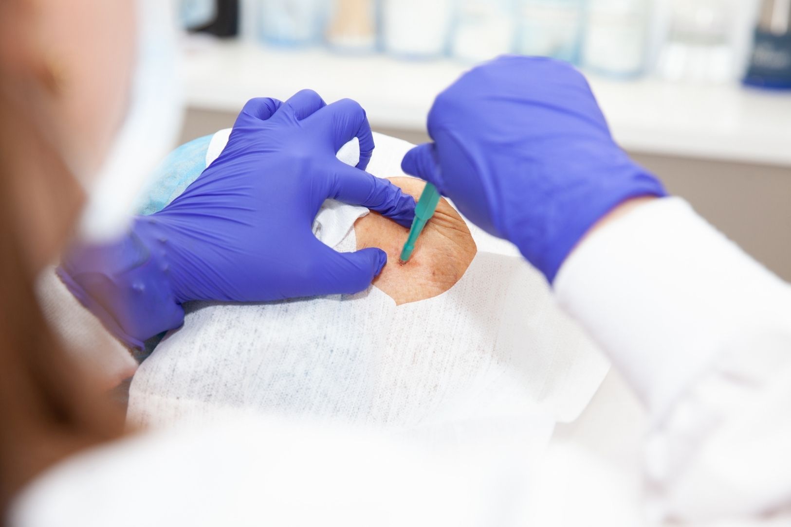
Skin biopsy is a diagnostic method that allows a small tissue sample to be taken from a diseased or suspicious area on the skin and examined under a microscope. It is used in the diagnosis of many diseases such as eczema, psoriasis, skin infections, and benign or malignant skin tumors. The procedure performed under local anesthesia is short, and the sample taken is evaluated in the pathology laboratory. Appropriate treatment is planned according to the results.
Why Is a Skin Biopsy Necessary?
A skin biopsy is performed to eliminate uncertainty and establish the most accurate treatment plan. Sometimes a lesion does not clearly say “this is the disease” even when examined by an experienced eye or with a dermoscope (illuminated magnifier). At this point, it is necessary to see the cellular structure of the tissue, in other words, its internal structure.
The main reasons why your doctor may request a skin biopsy are:
- Suspicion of cancer (Melanoma, Basal Cell, Squamous Cell)
- Precancerous lesions (such as Actinic Keratosis)
- Evaluation of atypical (suspicious) moles
- Psoriasis
- Lichen Planus
- Autoimmune diseases such as Lupus Erythematosus
- Pemphigus or Bullous Pemphigoid (blistering diseases)
- Chronic rashes that cannot be diagnosed
- Skin reactions that do not respond to treatment
- Some suspicious fungal or bacterial infections
What Information Should I Give My Doctor Before a Skin Biopsy?
Although this procedure is a minor surgical intervention, you must share certain information for your safety. Your health history is very important in planning the procedure and minimizing possible risks.
Especially medications and supplements that may create a tendency to bleed are of vital importance.
Some common medications and supplements that may increase the risk of bleeding:
- Aspirin
- Clopidogrel (Plavix)
- Warfarin (Coumadin)
- New-generation blood thinners (Rivaroxaban, Apixaban, etc.)
- Heparin (blood-thinning injections)
- Fish oil (High-dose Omega-3)
- Vitamin E supplements
- Ginseng
- Garlic (as a high-dose supplement)
You should not stop these medications on your own, but you must definitely inform your doctor. Your doctor will make a plan according to the importance of the medication and the type of biopsy to be performed.
You should also state all known allergies.
Allergies that you must definitely mention:
- To local anesthetics (Lidocaine, Prilocaine, etc.)
- To adhesive bandages
- To plasters (adhesive tapes)
- To antiseptic solutions (such as Iodine, Batikon)
- To antibiotic creams (especially those containing Neomycin)
Apart from these, if you have an inherited bleeding disorder such as hemophilia, if you have a pacemaker, or if you are pregnant, you must definitely share this information before the procedure.
Does Skin Biopsy Legally Require “Informed Consent”?
Yes, it definitely does. Informed consent is much more than simply signing a paper; it is a legal and ethical information process that takes place between you and your doctor. It means that you are comprehensively informed about the process before giving consent to the procedure to be performed.
What needs to be explained to you during this consent process:
- The purpose of the procedure (why it is being done and what is being investigated)
- How the procedure will be performed
- The possible risks of the procedure (such as bleeding, infection, scarring)
- Whether there are alternative diagnostic methods
- The possible medical consequences if you refuse to have the procedure
- That the sample taken will be sent to pathology for diagnosis
This process is an opportunity for you to ask all your questions and approve the procedure with no doubts in your mind.
What Are the Different Skin Biopsy Methods?
Which technique will be used is a strategic decision determined according to the type of lesion, the suspected diagnosis, its depth, and its location on your skin. The goal is always to obtain the best cosmetic result while providing sufficient tissue for the pathologist to make the most accurate diagnosis.
- Shave Biopsy
This is the most superficial biopsy method. It generally involves “shaving” the lesion off the surface of the skin using a scalpel or a special hand blade. You can think of it as taking a raised structure on the surface of the skin like a thin leaf. It usually does not require stitches, and bleeding is stopped with the help of a small cautery device. It is especially preferred in aesthetically sensitive areas such as the face and in superficial lesions (seborrheic keratosis, some superficial cancer types).
- Punch Biopsy
This is one of the most commonly used methods. A very sharp-tipped, circular, pen-like instrument (punch) is used. This instrument is pressed onto the skin at a right angle and rotated to remove a full cylindrical sample from all layers of the skin (epidermis, dermis, and sometimes down to the underlying fatty tissue). You can think of it like a paper punch or a small cookie cutter. The 3 mm and 4 mm diameter ones are most commonly used. It is especially ideal in the diagnosis of rashes (such as psoriasis, lichen), because it allows the pathologist to see all layers of the skin together. The wound is usually closed with one or two small stitches.
- Excisional Biopsy (Complete Removal)
In this method, the entire lesion is removed together with a small amount of surrounding healthy skin tissue (intact surgical margin), usually 1–3 mm. The sample extends down to the fatty tissue beneath the skin. This procedure serves both diagnostic and therapeutic purposes. It is the preferred method especially in small suspicious moles or lesions with a high suspicion of skin cancer (especially melanoma). Since the entire lesion is removed, the pathologist can also evaluate whether the margins are clear. The wound is closed with stitches.
- Incisional Biopsy (Taking a Portion)
In this method, not the entire lesion but only a representative portion (a slice) of it is taken. This method is generally chosen if the lesion is very large (for example, if it covers an area as large as the palm of a hand) or if it is located in a very cosmetically or functionally sensitive area such as the face, finger, or palm, and removing the entire lesion at the first stage would cause a major deformity. The aim is first to make the diagnosis and plan the treatment later.
Which Skin Biopsy Technique Is Chosen in Which Situation?
Your dermatologist makes this choice according to the diagnosis they suspect:
- Superficial Raised Lesions (Wart, Skin Tag, Seborrheic Keratosis): Shave Biopsy is generally sufficient. It is fast and the cosmetic result is usually excellent.
- Rash-like and Inflammatory Diseases (Psoriasis, Eczema, Lichen): Punch Biopsy is almost always preferred. The pathologist needs a full-thickness sample to see how the disease affects the layers of the skin:
- Suspicious Moles and Suspicion of Melanoma: The rule is to remove the lesion with Excisional Biopsy (completely). The reasons for this are very critical and should be discussed under a separate heading.
- Very Large Tumors or Sensitive Areas: Incisional Biopsy (taking a portion) may be more appropriate for diagnosis.
Contact Us for Anything You Wonder About Skin Health!
Why Is Skin Biopsy Different in Suspected Melanoma?
The rules are very strict for lesions suspected of melanoma. If a lesion carries suspicion of melanoma clinically and dermoscopically, the lesion must be removed completely (excisional biopsy) whenever possible.
The reason for this is: the most important factor that determines the course and treatment of melanoma is the “Breslow Thickness.” This is a millimetric measurement showing how far the tumor has progressed from the uppermost layer of the skin to the deepest point. In order for the pathologist to make this measurement accurately, they need to see the entire lesion and its deepest point.
If only a part is taken from a mole suspected of melanoma with a punch or if the top is removed with a shave biopsy, this can result in disaster. The piece taken may not represent the thickest part of the lesion, and while the pathologist says “0.5 mm thickness,” there may actually be “2 mm thickness” in another part of the lesion. This may lead to incorrect staging of the disease (understaging), incomplete treatment, and the patient missing an important stage such as “Sentinel Lymph Node Biopsy” (sampling from the arm).
Are there no exceptions? Of course there are. Sometimes, on the face or finger, in a very large lesion, an incisional (partial) biopsy may necessarily be taken, but this must be done by taking a full-thickness sample from the thickest and most suspicious part of the lesion.
Where Is Skin Biopsy Taken From in Rash-like Diseases?
Lesion selection is also critical for the diagnosis of rash-like (inflammatory) diseases. It is necessary to choose the “most correct” rash for punch biopsy.
The rule here is to find the “freshest” lesion.
A newly appeared, active, “angry-looking” rash should be preferred.
Lesions that have been scratched excessively and turned into wounds (excoriated) are avoided. Because scratching distorts the original tissue and may mislead the pathologist.
Very old, “faded,” pale lesions are also not preferred. Because in these lesions, the active signs of the disease may have diminished and the diagnostic findings may have disappeared.
Why Is a Special Skin Biopsy Required in Blistering Diseases? (DIF)
When autoimmune diseases characterized by blistering of the skin such as Pemphigus and Bullous Pemphigoid are suspected, in addition to routine biopsy, a special test called “Direct Immunofluorescence” (DIF) is required. This test detects the proteins (antibodies) of the immune system that attach to the skin and cause the disease.
The success of the DIF test depends entirely on taking the sample from the correct place, and this is one of the most common mistakes.
For DIF, the sample is not taken from the blister itself. Because the inside of the blister is inflamed and the proteins being searched for may be degraded there. This leads to a “false negative” result (as if the disease were absent when it is actually present).
The correct area is this: The sample should be taken from normal-looking, intact, or slightly reddened skin immediately next to an active blister (3 mm to 10 mm away). This is the place where the “war” has just begun and where the antibodies can be seen most clearly. In addition, this sample is NEVER placed in formalin; it is placed in a special transport fluid called “Michels medium.”
Is the Skin Biopsy Procedure Painful?
This is what patients are most curious about. Since the procedure is performed under local anesthesia, it is not painful. Patients generally feel only a mild “bee sting” or “burning” sensation lasting about 10–15 seconds when a very fine needle is inserted while the anesthetic substance is being administered.
After the area becomes numb (which happens within seconds), you do not feel any of the cutting, removal, or stitching procedures. Local anesthetics containing adrenaline (epinephrine) are generally preferred. Adrenaline has two important functions: it both prolongs the painless duration of the procedure and minimizes bleeding by constricting the vessels in the area.
How Should Wound Care Be After a Skin Biopsy?
Wound care after a biopsy is very important to prevent infection and minimize scarring. The basic principle of modern wound care is to provide “moist wound healing.” The wound drying out and forming a scab (desiccation) slows the movement of healing cells and delays healing.
Your daily care routine should be as follows:
- Wash gently with soap and water
- Rinse thoroughly (soap residue may cause irritation)
- Apply the ointment recommended by your doctor (usually simple petroleum jelly or a neutral moisturizer)
- Cover with a clean bandage
You will be asked to repeat this procedure every day until the stitches are removed or the wound is completely closed.
Which Mistakes Should Be Avoided in Wound Care?
Some common practices done while trying to speed up healing actually slow the process or create risk.
Some common mistakes that slow healing or create risk:
- Using oxygenated water (Hydrogen Peroxide)
- Applying alcohol or cologne
- Applying tincture of iodine (Iodine) on the wound
- Applying over-the-counter antibiotic creams (Neosporin, Bacitracin, etc.)
- Leaving the wound open so it can “air out” and waiting for it to dry
- Picking the scab off the wound
Especially oxygenated water and alcohol damage the delicate healing cells in the wound bed (they are cytotoxic). Over-the-counter antibiotic creams, on the other hand, are unnecessary and carry a high risk of causing “allergic contact dermatitis” (contact eczema). This may lead to the wound site being thought to be infected, more cream being used, and the problem growing.
Contact Us for Anything You Wonder About Skin Health!
Which Situations Are Urgent After a Skin Biopsy?
Complications are rare, but it is important to know what to do if they occur. There are two main urgent situations: uncontrollable bleeding and infection.
Bleeding: After you go home, the wound site may ooze. If active bleeding starts, apply firm pressure continuously to the area with a clean gauze pad for at least 20 minutes. Opening it to check every 2–3 minutes in order to control the blood is the most common mistake, because it disrupts the clot that is forming. Look at your clock and do not lift the pressure for 20 minutes. If at the end of this period the bleeding is still bright and actively continuing, you should call your doctor.
Infection: Infection generally shows symptoms 2–3 days after the procedure.
Signs of infection are:
- Increasing and persistent pain
- Expanding redness and swelling around the wound
- A noticeable increase in warmth in the area
- Purulent (yellow-green, foul-smelling) discharge from the wound
- Fever or chills
If any of these symptoms are present, you should consult your doctor immediately.
Will There Be a Scar After a Skin Biopsy?
It is necessary to be honest about this: Every full-thickness procedure that goes down to the dermis layer (middle layer) of the skin leaves some degree of scar. While shave biopsies may sometimes heal with only a color change, especially punch and excisional biopsies will definitely leave a permanent scar.
The goal here is not for the scar to be “zero” but for it to remain as little and as aesthetically as possible. For this reason, dermatologists plan the direction of the incision parallel to the natural tension lines (RSTL) of the skin. This allows the wound to heal with less tension and as a finer line.
The most important thing you should do so that the scar has the best long-term appearance is to protect the area from the sun. A fresh scar (the first 6–12 months) is extremely sensitive to the sun. Sun exposure causes the scar to darken permanently (post-inflammatory hyperpigmentation). Therefore, even after the wound has healed, it is essential to apply a high-factor sunscreen of at least SPF 30 to the area.
What Happens to the Skin Biopsy Sample Taken?
That small piece of tissue taken is the most important step for you. That piece is placed into special liquids called “fixative” or “transport medium.”
- Routine Examination: Most biopsies are placed in a solution of “%10 Neutral Buffered Formalin,” which “fixes” the tissue and prevents it from deteriorating.
- DIF Test: The sample taken for blistering diseases is NEVER placed in formalin. It is placed in a special transport fluid (Michels medium).
- Genetic Tests: Sometimes, for lymphoma or special cancer investigations, the sample is placed fresh or in special containers to be frozen (fresh-frozen).
In the laboratory, this tissue is cut into slices “thinner than a strand of hair,” stained with special dyes, and examined under the microscope by a pathologist. The pathologist writes a report by looking at the shape of the cells, their arrangement, and abnormalities in the layers of the skin. This report becomes the roadmap for your diagnosis and therefore your treatment.
Frequently Asked Questions
In which cases is a skin biopsy performed?
A skin biopsy is performed to diagnose moles, rashes, wounds, or suspicious lesions on the skin. It is especially preferred to confirm the diagnosis in diseases such as skin cancer, lupus, psoriasis, or eczema.
What should be considered before a skin biopsy?
Before the procedure, blood-thinning medications should be discontinued under a doctor’s supervision, and any allergies should be reported. It is important for the area to be clean, and there is no need to fast since local anesthesia will be applied.
How is a skin biopsy performed and how long does it take?
A skin biopsy is performed by taking a small piece of tissue under local anesthesia. The procedure generally takes 10-15 minutes, and the sample taken is sent to the laboratory for pathological examination.
What is the recovery process like after a skin biopsy?
After the procedure, there may be mild redness, swelling, or crusting in the area. It generally heals within a few days. Following the dressing and wound care instructions recommended by the doctor speeds up healing.
Will there be a scar after a skin biopsy?
After a biopsy, a very small scar may generally remain. The appearance of the scar depends on the amount of tissue taken and the person’s skin structure. When appropriate wound care is performed, the scar is generally not noticeable.
How long does it take for skin biopsy results to come out?
Depending on the laboratory workload, skin biopsy results are generally available within 7-10 days. When some special tests need to be performed, this period may be slightly longer.
Is a skin biopsy a painful procedure?
Since local anesthesia is applied during the procedure, no pain is felt. After the effect of the anesthesia wears off, there may be a slight ache, but it can generally be controlled with simple painkillers.
Can a skin biopsy be performed during pregnancy?
Yes, if deemed necessary, a skin biopsy can also be performed during pregnancy. The local anesthetics used are safe during pregnancy, but the doctor makes an evaluation before the procedure according to the week of pregnancy.
Is there a risk of infection after a biopsy?
As with every surgical procedure, there is a slight risk of infection after a biopsy. Following cleanliness rules, doing the dressing regularly, and following the doctor’s recommendations minimize this risk.
How is treatment planned according to the skin biopsy result?
According to the pathology result, the diagnosis becomes clear and the treatment is arranged accordingly. If cancer or a serious skin disease is detected, surgical intervention or drug treatment may be planned.